")
")
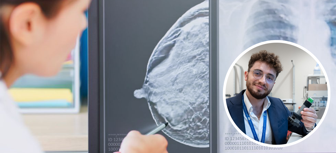
Breast cancer is among the most frequent neoplasms in terms of incidence in the female population and among the main causes of death in women worldwide.
It is estimated that one in eight women will be affected in their lifetime. Identification is crucial to increase the chances of recovery and survival from the disease.
Breast palpation is one of the most popular techniques for detecting tissue abnormalities in clinical scenarios, including breast examination. However, the tactile sensation used to identify tumors by the doctor or a woman during breast palpation makes this procedure subjective.
In recent decades, tactile sensors have been developed to quantitatively discriminate between cancerous and healthy tissues, but most of these systems still suffer from low sensitivity, high power consumption, reduced sterilization duration, and electrical noise.
Starting from these assumptions, a group of researchers in collaboration with the Breast Unit of the Campus Bio-Medico University Hospital Foundation, worked on the design of a device to overcome all these limitations, exploiting the advantages of fiber grid Bragg (FBG) technology combined with that of 3D to develop an innovative tactile probe for breast and cancer identification.
This device therefore exploits the advantages of the fiber Bragg grid (FBG) integrated into a 3D printed structure (Fig.1). The integration of FBGs during 3D printing combines the myriad advantages of fiber-optic technology (e.g., compactness, immunity to electromagnetic interference, non-electrical multiplexing capabilities, and high metrological shoring) with those of rapid prototyping (e.g., low-cost, high possibility of customization of the device to be printed, short manufacturing times).
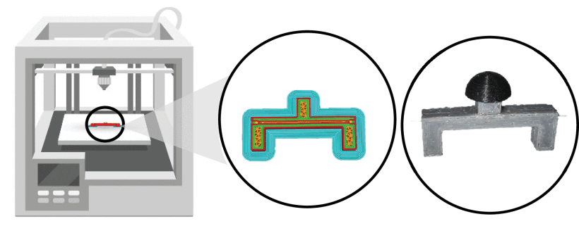
Fig.1
Finite element analysis and fiber placement optimization
Different types of analyses and tests were carried out for the design, starting with finite element analysis (FEA). To analyze the mechanical response of the designed structure when a load is applied to the top of the head, two finite element (FE) models were constructed. From the results obtained, it was noted that the distribution of the strain along the shear lines reached values such as to induce breakage of the FBG sensor. From these conclusions, it was considered to place the optical fiber along the blue cut line, as it ensures that the optical fiber is entirely in traction under load conditions, even if compression may occur if integrated into the base (Fig. 2). The sensor then works by feeling the surface of the breast to discriminate cancerous tissues from healthy tissues by detecting differences in stiffness.
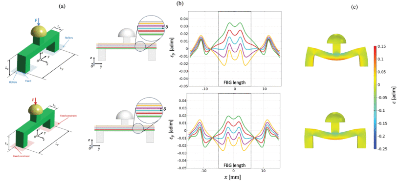
Fig.2
Structural design and geometric characteristics of the sensor unit
From these evaluations, the U-shaped structural sensing unit with a central column, the FBG sensor (1541 nm λB, 10 mm grating length and reflectivity >90%; marketed by AtGrating Technologies, Shenzhen, China) coated with silica and acrylate integrated into the structure was realized. The sensor head is adhered to the central column of the U-shaped structure with cyanoacrylate glue and as explained below, the integration of the FBG takes place during printing by inserting it into a grooved channel in the center of the tape. From the FE results, the sensor was designed to fit the base by integrating the flanges to a depth of 3mm.
All components of the detection unit were printed using fused deposition modeling (FDM) technology. The contact head has been molded in polylactic acid (PLA), a stiffer component of the U-shaped structure in which the sensor is placed, made of 95A TPU.
Sensor Unit Manufacturing Process
This ensured a complete transmission of the entire F applied to the head of the U-structure and, in turn, the FBG can be achieved. The main U-structure fabrication steps with sensor meshes are listed below:
- Pre-processing: Preparation of the g.code file of the computer-aided design using CURA slicing software, set a 100% infill density, triangular geometry infill and a print speed of 30 mm/s. Then, the job is sent to the 3D printer.
- Production: 3D printing phase. The fused filament (i.e. TPU 95A) is extruded from the nozzle and deposited layer by layer on the printer bed. At the 21st layer, the channel is created to incorporate the optical fiber. Printing is paused and the optical fiber is pre-tensioned in the channel. Printing is then restarted
- Post-processing: At the end of the process, the U-shaped structure that integrates the FBG during printing is removed from the bed, the PLA printed head is attached to the structure, and the sensor is ready to be inserted into the base.
Metrological characterization and testing on silicone phantoms
Subsequently, tests were carried out to detect the tactile Young(E) modulus on cylinders in two specific types of silicone rubber (e.g. Skin Drago 10A—ds10, more flexible and Skin Drago 30A—ds30, stiffer) and then evaluated the sensitivity capacity of the discriminant unit of materials with different stiffnesses. To recognize breast cancer from the surface of the body, the device should be able to discriminate tissues based on mechanical properties.
Tactile Probe Components: Puppet Design, Fabrication, and Testing
The probe consists primarily of multiple sensing units (e.g., three 3D-printed sensors based on FBG technology), a cover and base with a screw-threaded connection to protect the sensors, except for their contact heads, and a secured handle to allow the user easier application of pressure to the tissue (Fig. 3). Finally, a cap with three openings allowed the installation of the fiber optic LC adapters to allow the interconnection of the sensing units integrated into the tactile device to the FBG interrogator. The three detection units are located one in the center of the base and the others on the sides. As already described, a single FBG is embedded in each sensing unit along its neutral axis while the resting part of the optical fiber is first wound to the reel-like axis of the handle and then inserted through the handle to reach the adapter and couple the LC connector at the end of the fiber to that of the patch cords. All probe components are manufactured from PLA, except for the U-shaped structure of the sensing units, which is made of TPU.
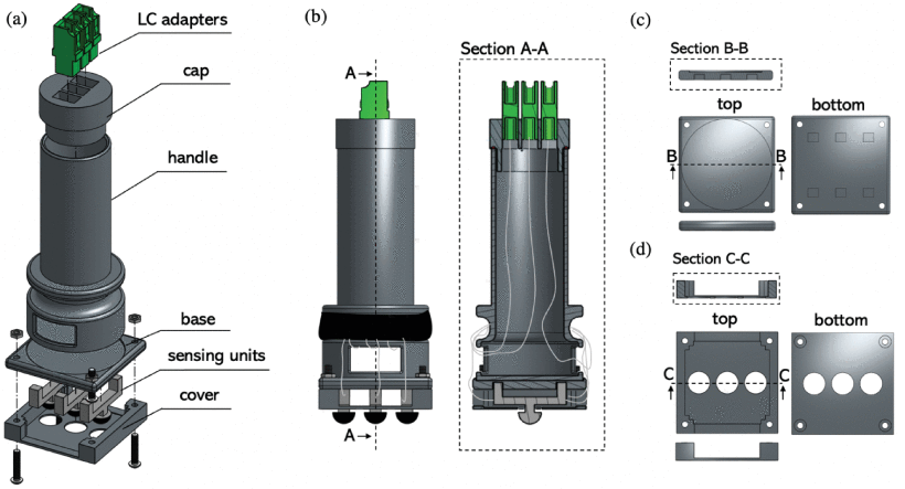
Fig.3
To better study the ability of the tactile probe to discriminate materials with different stiffness in the identification of breast cancer, a silicone phantom was developed that traces a model of breast with tumor lesion.
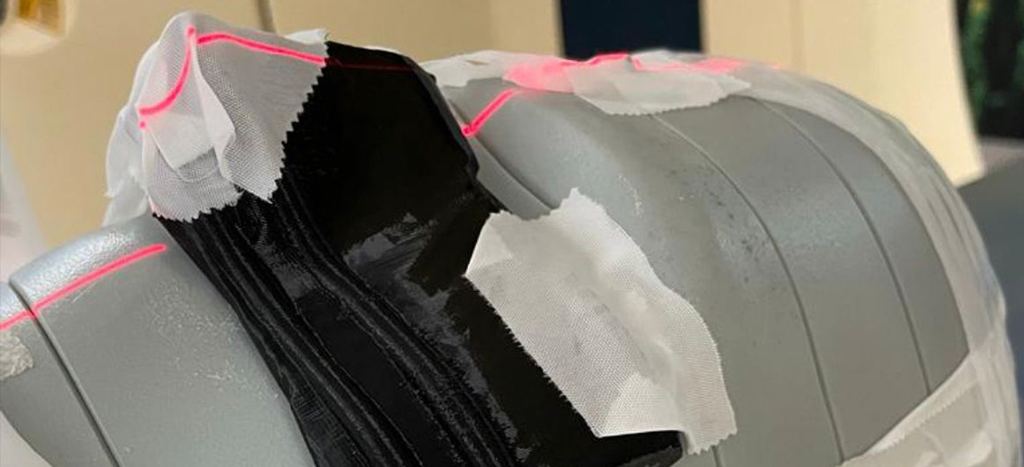
The test model was prepared by simulating breast tissue with ds10 silicone in which a 10mm diameter sphere 3D printed in PLA used to model the neoplastic tissue was dispersed (Fig. 4).
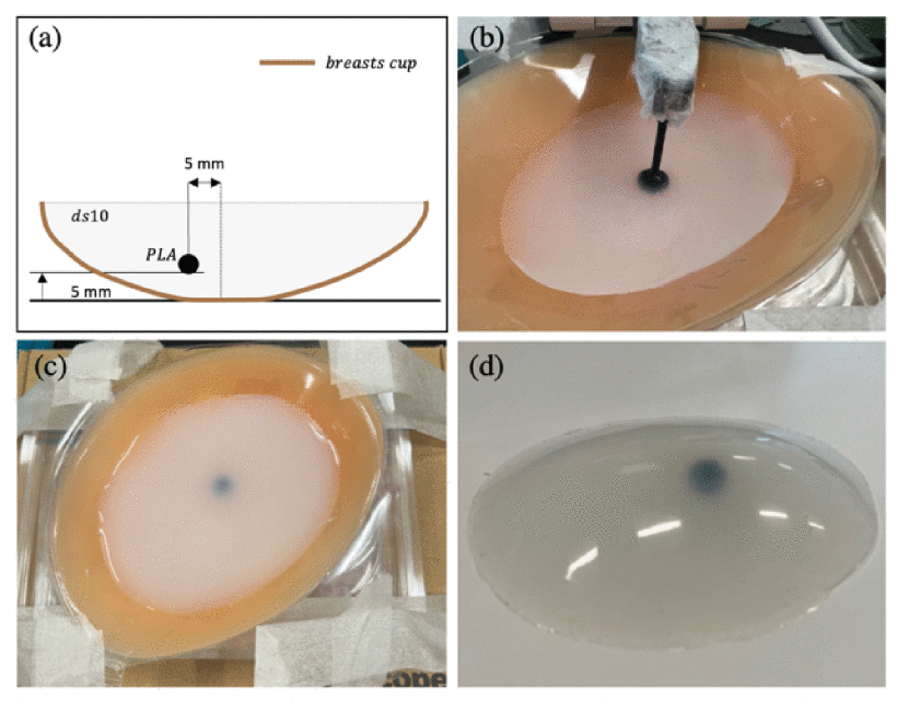
Fig.4
The ability of the tactile probe to detect the presence of neoplastic tissue inside the phantom was then analyzed using the compression machine already used for metrological evaluation. In these experiments, the handle of the tactile probe was locked by the upper grille and pushed against the phantom placed on the lower plate (Fig. 5)
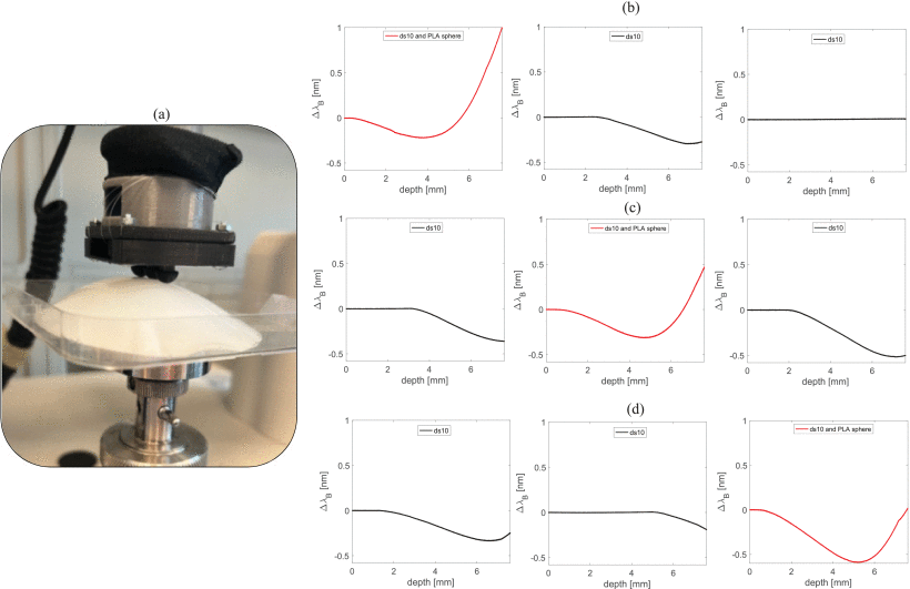
Fig.5
To date, only a few solutions have been marketed for palpation of breast tissue. The most used are silicone pads developed to reduce friction between the fingers and breasts and help women during self-examination, but there are no sensors integrated into the silicone matrix to automatically detect tissue abnormalities.
This device, by integrating the FBG into a 3D printed structure, makes the sensor more robust than detection solutions in silicone arrays and easier to sterilize than other devices in the literature. The use of FDM also plays a crucial role in the performance of the FBG sensor as better silicone adhesion is expected, and FEA-driven optimization can be easily adopted by customizing print settings and materials, thus reducing time and cost.
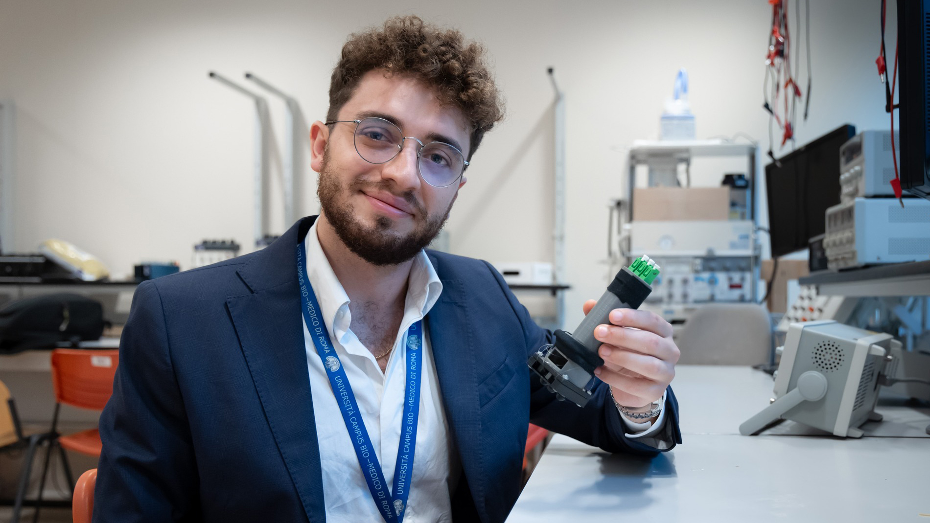
The project contained in the thesis work of Alfredo Dimo, research assistant at the Laboratory of Biomedical Measurements and Instrumentation UCBM has been recognized with the IEEE Italy Section Award - ABB 2023 Master Thesis Award Technology Category.
Thanks to these successes, the project is already working on the next optimizations and improvements, focused on increasing the spatial resolution of the probe to estimate the size and stiffness of the breast tissue nodule and the addition of a control unit to signal to the user when the threshold value is exceeded.
Source: IEEE.org